SynALI is a novel Air Liquid Interface model mimicking lung architecture. The microfluidic device is functionalized with epithelial cells surrounded by vasculature comprised of endothelial cells. The SynALI structure maintains an Air Liquid Interface across the airway cells. As a result, airway tubules form and transport mucus and are maintained by the surrounding epithelium. Cell morphology, airway structure, cell-cell interactions, and functions of the airway (e.g. mucus transport, ciliary beating, therapeutic-induced improvements) can be visualized and quantified in real-time in both normal and diseased conditions.

Unique Features include:

  • Morphologically realistic airway structure and environment
  • Air Liquid Interface (ALI) across the epithelium and endothelium
  • In-Vivo hemodynamic shear stress
  • Real-time visualization of cellular and barrier functionality including mucus, ciliary beating, immune cell interactions, and therapeutic screening
  • Robust and easy to use protocols
synali-apical
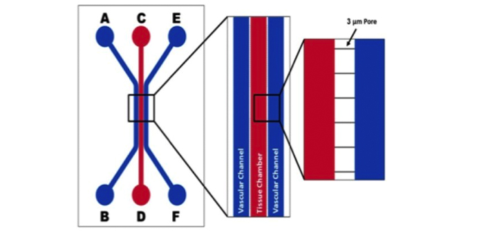
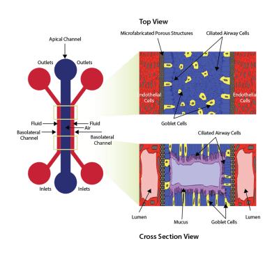
Schematic of the device used to develop the Air Liquid Interface across the endothelial and epithelial cells. The air (or epithelial) channel is separated from two-fluid (basolateral) channels by a microfabricated porous structure. The right panel shows the orientation of cells when seen from the top and cross-section view.

Examples of Models Developed Using SynALI Devices

synali small airway co-culture model
Small Airway lung model – Co-culture with human bronchial epithelial cells (HBEC) and lung microvascular endothelial cells .
synvivo synali alveolar lung model
Alveolar lung model with human microvascular lung endothelial cells and alveolar epithelial type I and II cells.

PRODUCT PURCHASING OPTIONS

Chips: Depending on your specific research applications you can select from IMN2 linear chip configurations.

Kits: All the basic components required to run SynALI assays can be purchased as kits. Two  formats are available:

Starter Kit: Select this for your first-time purchase

  • 10 SynALI chips -IMN2 linear (3um slits)
  • Accessories including tubing, clamps, needles, and syringes
  • Pneumatic priming device (required for priming tubing to remove air)

Assay Kit: Select this kit format if you have previously purchased the pneumatic priming device

  • 10 SynALI chips – IMN2 linear (3um slits)
  • Accessories including tubing, clamps, needles, and syringes
     (Kits do not include the air pump needed to establish the air-liquid interface). 

IMN2 Linear

SynBBB IMN2 linear
Idealized Co-Culture Network (IMN2 Linear) Chip: 3 um slits. Channel Width; 200-500-200um,  50um Travel (distance between channels), 100um Depth (height). Cat# 108011

Starter Kit

$1,700Add to cart

Assay Kit

$1,500Add to cart

SynVivo Platform Used to Create Human Airway-On-Chip

Researchers from the University of Alabama, Birmingham report on the use of the SynVivo platform for the development of a human airway-on-a chip model. Combined with novel micro-optical coherence tomography (µOCT), this model enables non-invasive quantitative imaging of ciliary movement. The imaging includes beat frequency and mucociliary transport.

“The advantage of this microfluidics device lies in the formation of a complete lumen for both the airway epithelium and the adjacent endothelium. It is a step forward in the development of a model that recapitulates both the cellular differentiation and organization into tubular structures, similar to the small airways and microvasculature”, said Dr. Jennifer Guimbellot, pediatric pulmonologist and assistant professor, UAB School of medicine.

The airway model was developed with a co-culture of primary epithelial cells and endothelial cells across an Air Liquid Interface (ALI). It was created using a customized SynVivo microfluidic chip enabling real-time quantitative imaging. The functionality of the developed airway-on-a-chip model was demonstrated by monitoring active cilia, mucus-producing cells, and biomarkers of cellular function under physiological conditions.

Validation From Researchers

According to Dr. Steven Rowe, Professor of Medicine and Director of the Gregory Fleming James Cystic Fibrosis Research Center: “Developing new tools that appropriately model the intact mucociliary transport apparatus of humans is a major priority, and has implications for biological research of airway diseases including cystic fibrosis”. The developed airway model represents a new approach to personalized medicine and as a predictive tool for pharmaceutical development.

Co-cultured microfluidic model of the airway optimized for microscopy and micro-optical coherence tomography imaging
Authors: Zhongyu Liu, Stephen Mackay, Dylan M. Gordon, Justin D. Anderson, Dustin W. Haithcock, Charles J. Garson, Guillermo J. Tearney, George M. Solomon, Kapil Pant, Balabhaskar Prabhakarpandian, Steven M. Rowe, and Jennifer S. Guimbellot.
Biomedical Optics Express Vol. 10, Issue 10, pp. 5414-5430 (2019).
Epi-and-endothelial-co-culture-in-chip-ALI-500
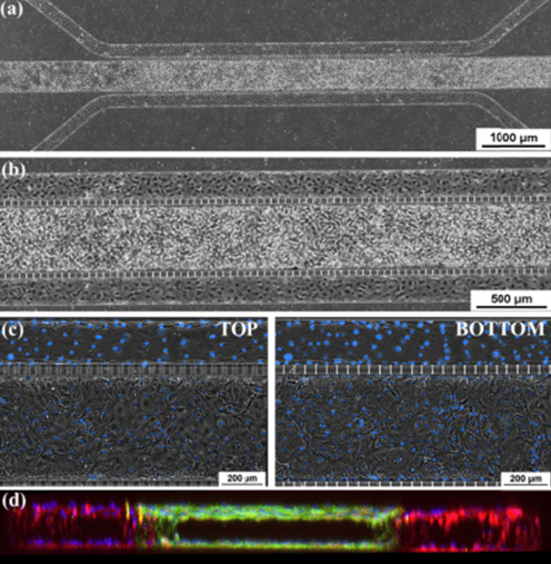
Epithelial and Endothelial Co-Culture in Microfluidic Chip. (a) and (b) Phase Contrast image of the confluent co-cultured chip with epithelial cells in the center channel and endothelial cells lining the peripheral channels. (c) Phase Contrast of top and bottom of the channel showing confluent monolayers in tissue and vascular channel. These images show the confluence of cells on both the top and bottom of the center channel and one side channel with a central, clear lumen. (d) Cross-sectional, 3-D reconstructed confocal image of co-cultured chip showing lumen formation in all three channels (10X mag.). Taken from Liu et al 2019.
ALI-differentiated-cell-staining-500
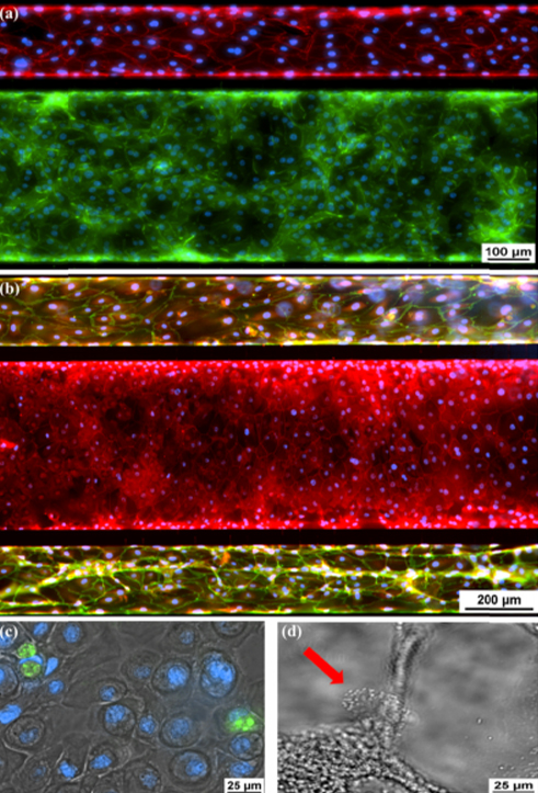
Immunofluorescence of Differentiated Epithelial and Endothelial Cells. (a) Ecadherin (green), epithelial cell adherin protein. VE-Cadherin (red), endothelial cell adherin protein. (b) ZO-1 (red) staining tight junctions of epithelial cells in central channel and also VE-Cadherin staining (green) in vascular channel. (c) Muc5ac (green), intracellular mucin staining inside of epithelial cells. (d) Ciliated epithelial cell indicated by the red arrow.

Small Airway Lung Model Exhibits Mucus Formation and Biomarker Staining

SynALI-mucus formation and biomarker staining
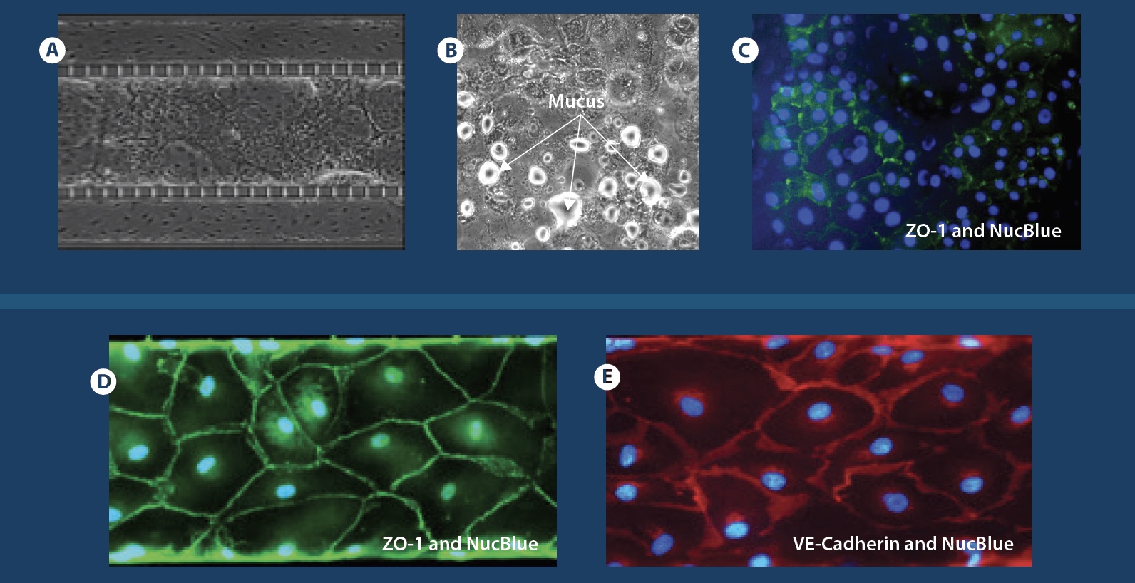
A-C Confluent co-culture of lung endothelial and epithelial cells following ALI development highlighting mucus formation and staining of biomarkers in epithelial cells. D-E Biomarker staining for tight junction markers (VE-Cadherin and ZO-1) in endothelial cells.

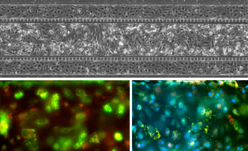
Alveolar Lung Model: Co-Culture and Tri-Culture Options

Co-Culture with biomarker staining

Alveolar-co-culture
Top image shows lung microvascular endothelial and epithelial cell co-culture in the SynALI devices. Bottom images: Red: Type I Cells: HT_56 antibody. Green: Type 2 Cells: HT_280 antibody. Blue: Nuclei

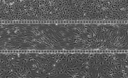
Tri-culture with Lung Endothelial Cells, Fibroblasts, Epithelial Cells

Alveolar-Tri-culture
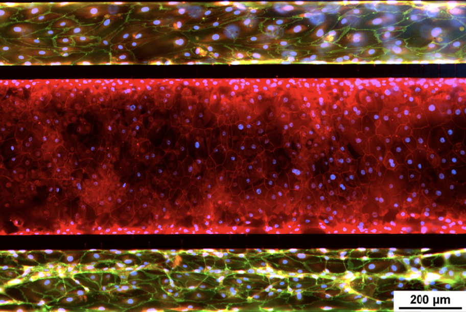
Phase images of SynALI Alveolar model tri-culture: Top channel: Lung microvascular Endothelial cells. Middle channel: Lung Fibroblasts. Bottom channel: Lung Epithelial cells.

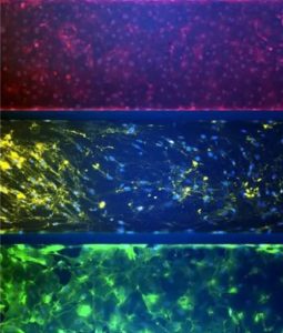
Biomarker Staining

SynALI Alveolar Model 500
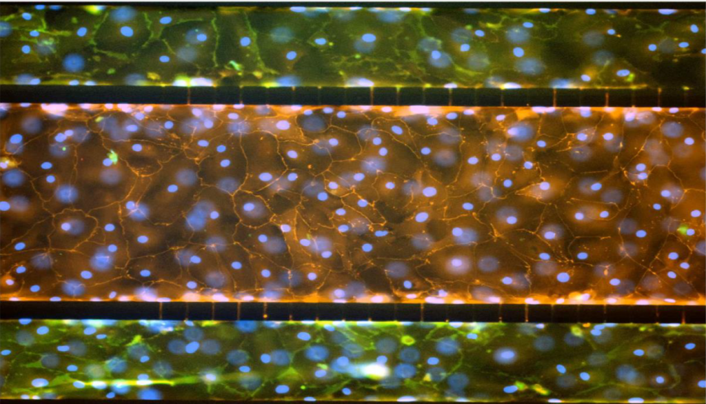
Triculture Alveolar lung model showing biomarker staining – VE-Cadherin and Nuc Blue in lung endothelial cells, Lung Fibroblast Surface protein and Nuc Blue in the middle fibroblast layer and  E-Cadherin, and Nuc Blue in the epithelial cell (bottom)

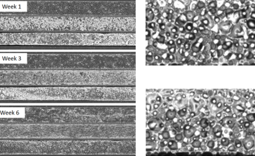
Long Term Tri-Culture and Mucus Secretion

Long-term-tri-culture-mucus-secretion
Alveolar Lung Model: Left image shows culture over weeks1 through week 6. Mucus secretion is shown on the right.

Assay Development and Screening using SynALI:

Models Available: 

  • Monoculture using primary epithelial cells
  • Co-Culture with endothelial cells
  • Tri-culture with fibroblasts

Assays:

  • Toxicity assays
  • Biomarker analysis
  • Therapeutic screening

Endpoints to choose from:

Vascular permeability using fluorescent-tagged molecule, ROS, biomarkers, collect cells or effluents for downstream genomic, proteomic, or transcriptomic analysis

Contact us for more details on using the SynALI model for your research applications!

Co-Cultured Microfluidic Model Of The Airway Optimized For Microscopy And Micro-Optical Coherence Tomography Imaging
Authors: Zhongyu Liu, Stephen Mackay, Dylan M. Gordon, Justin D. Anderson, Dustin W. Haithcock, Charles J. Garson, Guillermo J. Tearney, George M. Solomon, Kapil Pant, Balabhaskar Prabhakarpandian, Steven M. Rowe, and Jennifer S. Guimbellot.
Biomedical Optics Express Vol. 10, Issue 10, pp. 5414-5430 (2019).
Manuals For This Model:

SynALI Lung Model-BROCHURESYNALI 3D LUNG MODEL TECHNICAL MANUAL

SynALI 3D Lung Model – Starter Kits

Includes consumables (10 chips, 100ft tubing, 25 slide clamps, 50 blunt tip needles, and 50 1 ml syringes). Starter kits will also include the pneumatic priming device (required to prime the air out of the tubing).

Note: Kits do not include the air pump needed to establish the Air Liquid Interface or other consumables such as cells, media and matrix. Laboratory equipment required includes incubators, inverted microscopes, and syringe pumps.
Filters Sort results
Reset Apply
CAT#
Product Name
Price
405001
$1,700
Max:
Min: 1
Step: 1
ADD TO CART

SynALI 3D Lung Model – Assay Kits

Includes consumables (10 chips, 100ft tubing, 25 slide clamps, 50 blunt tip needles and 50 1 ml syringes).

Note: Kits do not include the air pump needed to establish the Air Liquid Interface or other consumables such as cells, media and matrix. Laboratory equipment required includes incubators, inverted microscopes, and syringe pumps.
Filters Sort results
Reset Apply
CAT#
Product Name
Price
405002
$1,500
Max:
Min: 1
Step: 1
ADD TO CART

SynALI 3D Lung Model – Chips

Filters Sort results
Reset Apply
CAT#
Product Name
Price
108013-3
$350
Max:
Min: 1
Step: 1
ADD TO CART
108011-SA3
$350
Max:
Min: 1
Step: 1
ADD TO CART